As a developmental psychology minor, the majority of my time is spent either interacting with
children or learning about how children develop and grow. A large portion of my
education in this field has been about the brain development that takes place
immediately after a baby is born, and in their first few years when children
begin learning about the world around them. As synaptic connections are formed
and lost, a child’s brain becomes full of neuronal connections that
ultimately influence their ability to learn language, visually perceive the world around them, engage
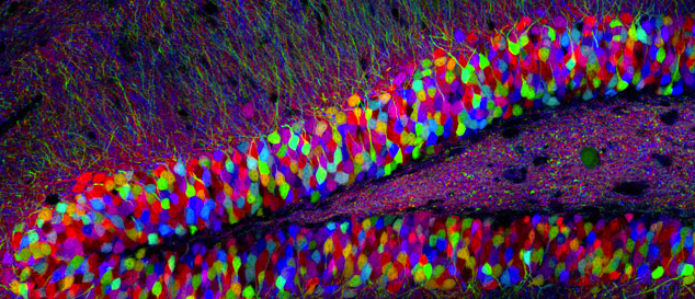
in higher level thinking, and much more. I found this week’s discussion of
Brainbow to be fascinating, as these neural connections can truly be visualized
to understand how synaptic pathways in the brain are formed.
 |
| https://littlesearcher.files.wordpress.com/2013/02/lichtmanlivet_hippocampus5x71.jpg |
Neurons in the brain communicate through synapses, which
are created both before birth and in the years after. Following puberty, however, some of these synapses
are lost during the process of synaptic pruning. Known as an “experience-dependent”
process, synaptic pruning depends strictly on an individual’s experiences. A true example of the "use it or lose it" phenomenon, synapses that are no longer used to transmit information between neurons are lost. Synaptic pruning can be exemplified in language acquisition, such that if a child is exposed to a language in their first few months of life, synaptic connections form in the brain that allow a child to differentiate between sounds of that particular language. If, however, that child never hears that language again, those synapses are pruned during or after puberty, making it far more difficult for the child to differentiate between sounds.
 |
| https://psychneuro.files.wordpress.com/2013/03/childs-brain1.jpg |
The Brainbow technique directly applies to early brain development, in that it allows scientists and researchers to view individual
neuronal pathways in the brain in a visually beautiful way. Each neuron can be distinguished from other
neurons with colorful fluorescent proteins that are used to flag particular
neurons with different colors. The colors used are derivatives of green
fluorescent proteins, that allow for the bright, colorful illumination of neural
connections in the brain. This mapping method allows scientists to trace the
pathways of neurons that connect different brain regions, allowing the
visualization of complete neuronal circuits.
 |
| http://i.imgur.com/cGtwE.jpg |
 |
| https://artscolab.files.wordpress.com/2012/12/tamily-weissman.png |
Works Cited
"Brainbow."
Center for Brain Science. N.p., n.d. Web. 17 May 2015.
<http://cbs.fas.harvard.edu/science/connectome-project/brainbow>.
Cai, Dawen, Kimberly Cohen, Tuanlian Luo, Jeffrey Lichtman, and Joshua Sanes. "Improved Tools for the Brainbow Toolbox." Nature (2013): n. pag. Print.
Galvan, Adriana. UCLA. Sept. 2014. Lecture.
"The 100 Colours of the Brainbow." Neurophilosophy. N.p., n.d. Web. 17 May 2015. <http://scienceblogs.com/neurophilosophy/2007/10/31/the-100-colours-of-the-brainbow/>.
Vesna, Victoria. “Neuroscience and Art.”
Cole UC online. Youtube, 15 May 2015. Lecture.
No comments:
Post a Comment